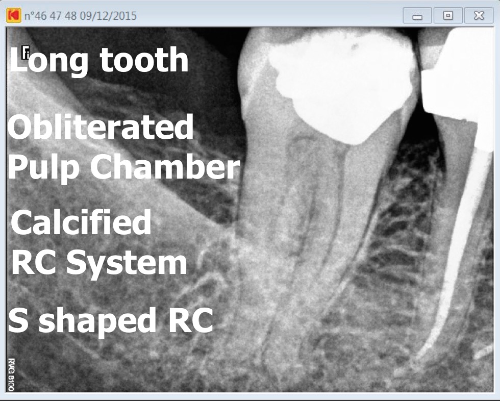
calcified pulp chamber
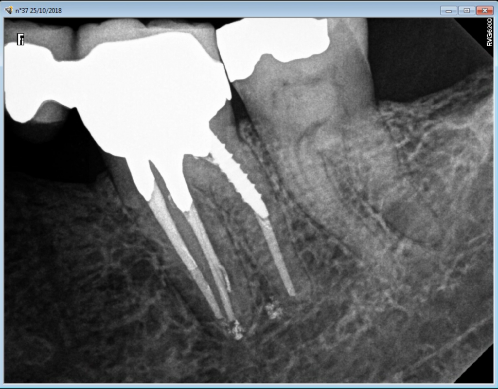
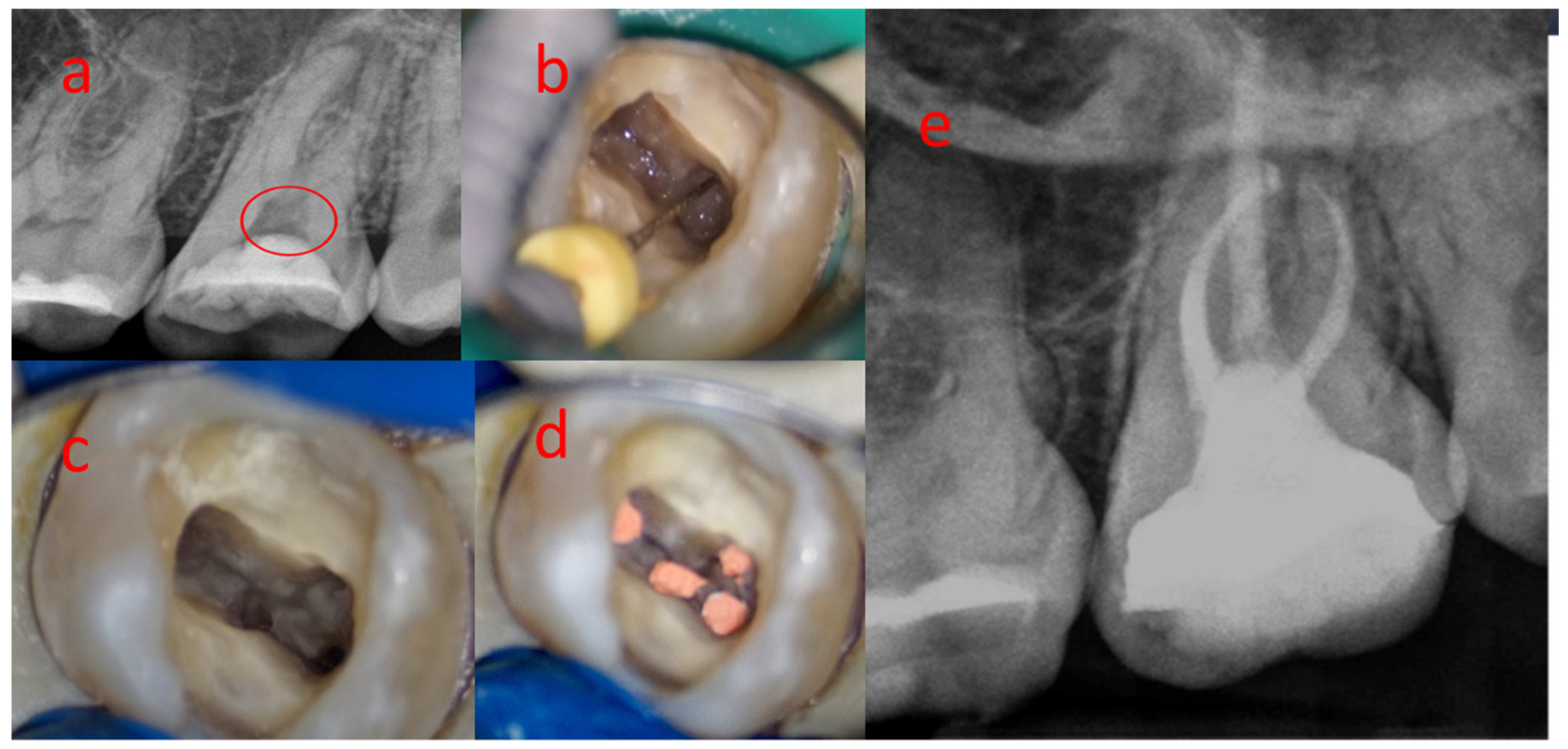
Root canal entries are also embedded. Concerning the ceiling of the pulp chamber two important facts were discovered.

Response To Pulp Sensibility Tests After Full Pulpotomy In Permanent Mandibular Teeth With Symptomatic Irreversible Pulpitis A Retrospective Data Analysis Journal Of Endodontics
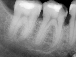
The diffuse or linear deposits are typically found in the root canals and generally are parallel to the blood vessels.

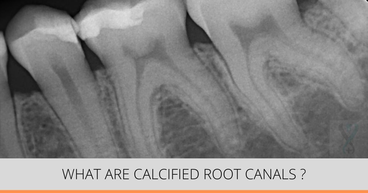
. The term calcified tooth means a tooths normal dental pulp chamber is compromised due to a reduction in size or obliterated due to trauma disease decay or age. Secondly we should be able to identify and. When a dental professional takes an X-ray of a tooth with calcification they may notice its abnormal appearance.
Primary dentin is the main. Thus we estimated the depth and got as close as possible to the pulp chamber roof. Based on morphology TRUE DENTICLES Localized masses of calcified tissue that resembles dentin Resembles more of secondary dentin More common in pulp chamber than in root canal Seldom larger than a fraction of millimeter usually located near the apical foramen.
The ceiling is at the level of the CEJ 98 of the time Figures 4a and 4b. FALSE DENTICLES Do not exhibit dentinal tubules Appear as lamellae deposited around a. Initiate the removal using straight and continuous movements over the calcification dry procedure do not irrigate.
The dental pulp or root canal system of a tooth is living tissue with a blood suppy and nerves and untold millions of cells. May be composed of irregular dentin true denticle or due to ectopic calcification of pulp tissue false denticle. Endō-lith A calcified body found in the pulp chamber of a tooth.
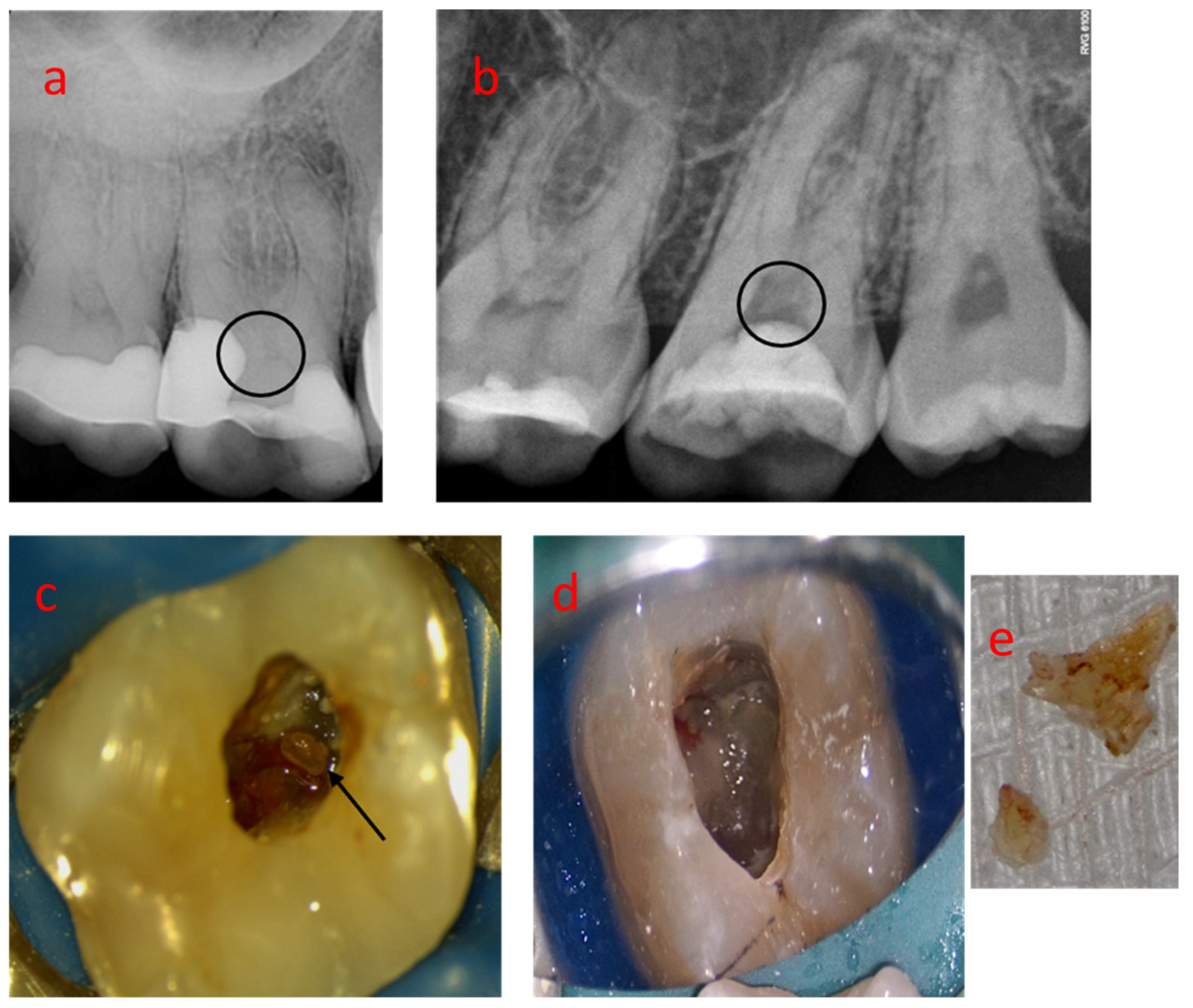
CAUSES CaOH2 is characterized by its ability to induce reparative bridge formation when applied to vital pulpal tissues However the pulp chamber and the pulp canal entrances can be subjected to dystrophic calcification after being exposed to CaOH2 for a long period It was suggested that the high alkaline pH level of CaOH2 irritates the pulp cells and activates. This pulp chamber calcification is caused by excessive dentin apposition by the odontoblasts that may be accelerated because of trauma to the tooth Abbott and Heah 2009. The maxillary molar endodontic access opening.
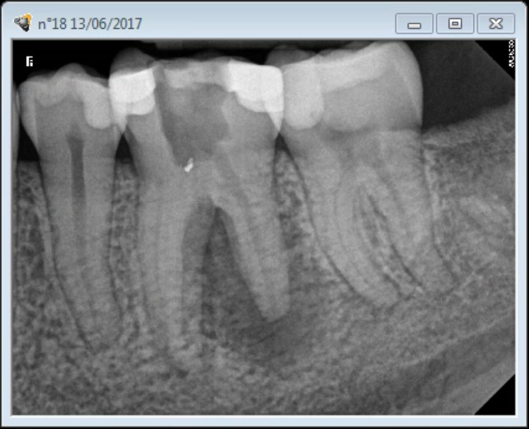
The root canal space should contain pulp but a calcified tooths chamber fills with hard calcified tissue. Root Canal - Calcified Pulp Chamber With a Guide. Root dentine the floor of the pulp chamber and calcified deposits.
After achieving access to the pulp chamber using a high speed turbine place the ultrasonic tip in direct contact with the pulp calcification. This 15-mm to 20-mm measurement is the most variable due to calcifications because of aging caries and restorations. Pulp chamber is almost completely obliterated by adherent calcifications.
The source is Endoacademie. The variation in incidence figures may be due to a number of factors such as the composition of the sample that was studied as well as the method of study and the restrictions. Pulp calcification may be of microscopic size or may be large enough to be detected radiographically.
This tissue is involved in the formation of teeth and cells in there are responsible for laying down the dentin the main body of your teeth that lies under the almost diamond-hard enamel. Consequently there is a decrease in the translucency of the tooth resulting in a. The height of a pulp chamber is between 15 to 20 mm Figure 5a.
This is especially true when the tooth being treated is heavily restored malposed or calcified. Locate more anatomy improving the quality of. Within the pulp chamber.
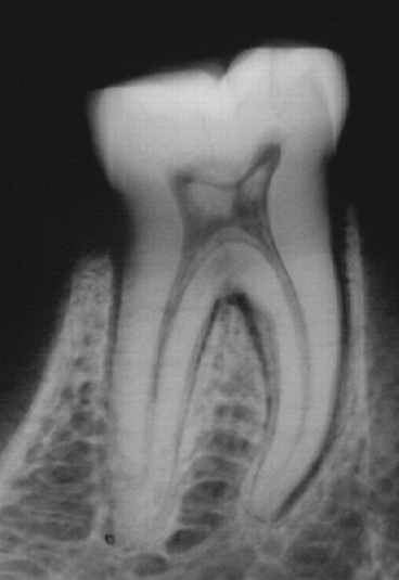
Pulp stones usually are found in the pulp chamber. A brief description of management of calcified pulp chamber and canals is given hereReferences-Mamoun JS. National Center for Biotechnology Information.
They often develop in teeth that appear quite normal in other respects. At this point the search for the distal canal begun as it was more coronally positioned. Pulp calcifications stones are nodular calcified masses appearing in either or both the coronal or root portions of the pulp organ.
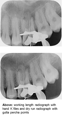
Microscope could not be of some use at the beginning of procedure to locate the root canal entries. Bordonne talks about guided endodontics. This article presents the endodontic management of a tooth with an obliterated pulp chamber and associated with a discharging sinus in a teenage patient.
Locating the number and position of orifices on pulp-chamber floors can be difficult. INCIDENCE Published figures of the occurrence of calcified structures in human dental pulps vary widely from a low of 75 to a high of 90 4 6 7 9-18 Table 1. The role of a calcium hydroxide lining to induce mineralization and cause the obliteration of.
In order to estimate the pulp chamber depth as well. Calcifications may be diffuse linear or nodular pulp stones. The canal itself may also appear significantly narrower or not visible because of the buildup of calcified tissue.
Denticle 1 pulp calcification pulp calculus pulp nodule pulp stone. They have been seen in both functional as well as embedded teeth. They are usually asymptomatic unless they impinge on the nerves or blood vessels.
When the first most coronal pulp chamber floor has been reached with a 330 long shank bur the root canal entries were still obscured by reparative dentine and could not be located despite the high magnification OPMI PRO ergo from Zeiss. As an IJHS review explains the X-ray may show that the pulp chamber is either hardly visible or not visible at all.

Types Of Pulp Diseases Dental Pulp Exposure Reversible Pulpitis Dental Pulp Calcification Irreversible Pu Teeth Diagram Sensitive Teeth Human Teeth
Dental Pulp Calcification And Changes With Age Bite Point

Dental Pulp Calcification All The Causes Bauer Smiles
Pulp Stones Friend Or Foe

Tooth Morphology Basics Teeth Dental Terminology Teeth Covers
Pulp Stones
Obliterated Root Canal System Endomontreal
Pulp Obliteration And Root Canal Endomontreal
What Are Calcified Or Blocked Root Canals Expert Dental Care
Medicina Free Full Text The Pulp Stones Morphological Analysis In Scanning Electron Microscopy And Spectroscopic Chemical Quantification Html
Clinical Tips For Instrumenting Calcified Canals Dentistry Today
Dental Pulp Calcification And Changes With Age Bite Point

An Access Is Set Up To Make A Smooth Straight Line Way To The Canal Framework And The Apex When It Is Effectively Done It Dental Health Dental Hygiene Dental
Pulp Stone Observed Inside The Pulp Chambers Of The Molars And Download Scientific Diagram
Saving A Severely Calcified Tooth With A Root Canal Procedure Endomontreal
Root Canal Procedure On A Calcified Second Molar Endomontreal
Medicina Free Full Text The Pulp Stones Morphological Analysis In Scanning Electron Microscopy And Spectroscopic Chemical Quantification Html

Endodontic Management Of A Tooth With Large Pulp Stone Style Italiano Endodontics

Identifying Pulpal Necrosis For Endodontic Treatment Spear Education
0 Response to "calcified pulp chamber"
Post a Comment